Treating AVN requires active intervention to slow down its quick progression and provide immediate and, ideally, long-term relief for the patient. Avascular Necrosis is an aggressive condition, and the health and resilience of the impacted joint will determine which treatment options are available.
Once a diagnosis is reached, your doctor will recommend the use of non-steroidal anti-inflammatory medications, plenty of rest, limited use of the affected joint, ice, compression, and elevation (if applicable) to reduce excessive swelling and inflammation. If it is determined that your AVN is pre-collapse, you are eligible for core decompression, free vascularized bone graft, and free periosteal core decompression (FPCD). For post-collapse patients, joint replacement surgery will most likely be the recommended treatment plan.
We’ll explore each of these treatment methods and discuss why we recommend FPCD for eligible patients.
Free Periosteal Core Decompression
Free periosteal core decompression is the method of treatment utilized at IFAR for its superior effects on AVN patients. It’s an enhanced approach to the standard core decompression procedure that accounts for the long-term health of the affected bone tissue. Just as in core decompression, a hole will be drilled into the suffering bone tissue to allow the surgeon to remove dead bone. This creates space, relieving pressure on the joint and potentially improving mobility.
The next step in FPCD, is to harvest healthy bone tissue (periosteum) from the patient, which will be utilized to protect the newly structured joint and reintroduce blood supply to the dying areas of the bone. This step is vital in the recovery process. The only alternative to regressing the development of AVN would be temporary methods (core decompression and medications), or total joint replacement.
Through the hole created by the core decompression procedure, the surgeon inserts the newly harvested periosteum in the precise location to allow the tissue to cover the affected bone and connect with the existing veins in the joint. This more completely addresses the root cause of poor blood flow that degraded the health of the original bone tissue.
Free Vascularized Bone Graft
Free vascularized bone graft for AVN involves the same procedure of core decompression, with the additional process of harvesting a graft of the patient’s bone, which is then placed into the affected joint for improved stability. Additionally, a bone graft that is vascularized rather than nonvascularized will introduce blood flow to the newly structured joint. This decreases the likelihood of the body rejecting the graft and, since AVN is caused by a lack of blood supply, is particularly relevant for eligible osteonecrosis patients.
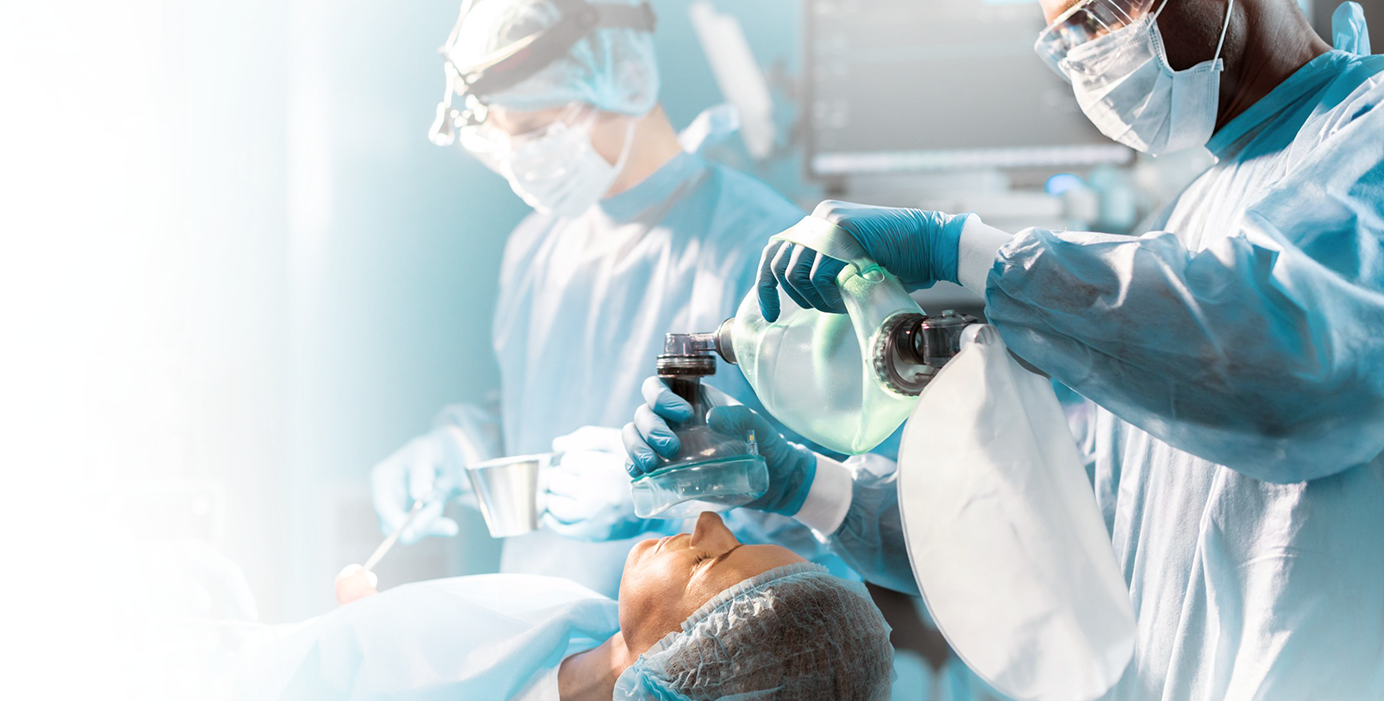
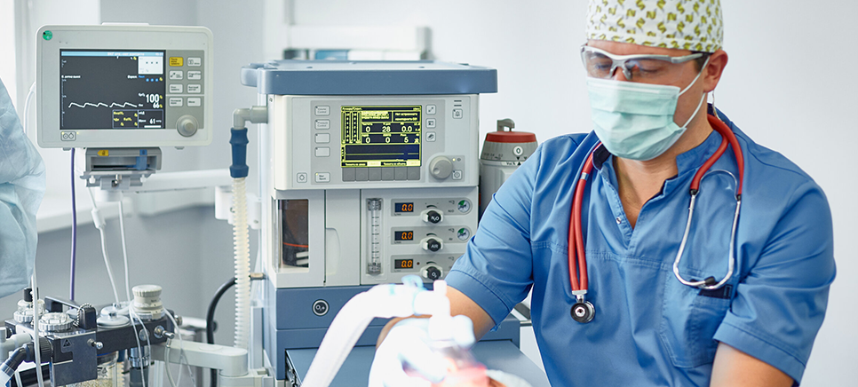
This procedure is done under general anesthesia, and patients will go home on the same day of the operation. The recovery period for free vascularized bone grafts depends on the type of joint operated on, the patient’s age, and a few other factors. Most patients will experience a near full recovery in six months following the procedure. Follow-up appointments will be set to monitor the progress of healing, physical therapy exercises will be introduced, and annual follow-up appointments may be recommended indefinitely.
Free Periosteal Core Decompression (FPCD)
Free periosteal core decompression is a very similar procedure to free vascularized bone graft, with the key difference being the use of periosteal tissue rather than bone. The periosteum is the outer membrane that coats the body’s bone tissue. By using periosteum, the procedure is less invasive than a bone graft while still introducing healthy, vascularized tissue into the affected joint. This provides immediate relief and long-term stability for the joint.
This is a newer treatment for AVN and one that very few surgeons have the knowledge and capability to perform. Fortunately, Dr. Adam Saad at IFAR specializes in this complex microsurgery, which has shown to provide exceptional relief for pre-collapse osteonecrosis patients, with minimal effects on the patient and just a three-month recovery period.
This procedure is done under general anesthesia, and patients typically go home on the same day of the operation.
Recovery from FPCD
Free periosteal core decompression is not only highly effective in improving the condition of osteonecrotic joints, but it is a shorter surgery and recovery period for the patient as well. FPCD is a shorter surgery than a free vascularized bone graft. This is credited to the difference in the invasiveness of harvesting periosteum versus grafting a patient’s bone tissue.
Patients will be able to start load bearing six weeks after the procedure and should make a full recovery in just three months following the operation. Patients typically experience less complications from harvesting periosteum compared to grafting a large section of bone, which can create nerve damage and chronic pain. FPCD is also more reliable than total joint replacement in that it does not use foreign materials. This creates less opportunity for infection or for any materials used in the procedure to fail or be recalled.
Patients will receive all relevant continued care information following the procedure. Follow up appointments will be set to ensure your recovery is on schedule and free from unnecessary setbacks. If at any time after the procedure you have any questions, you can always reach out to the IFAR team for answers or to schedule an appointment.


